In a significant stride towards democratizing advanced imaging techniques, a team of researchers from University College London, led by Dr. Silvia Cipiccia, has successfully demonstrated the potential of laboratory X-ray sources for high-resolution, quantitative phase nano-imaging. This development could bring powerful imaging capabilities typically reserved for large-scale synchrotron facilities to more accessible laboratory settings.
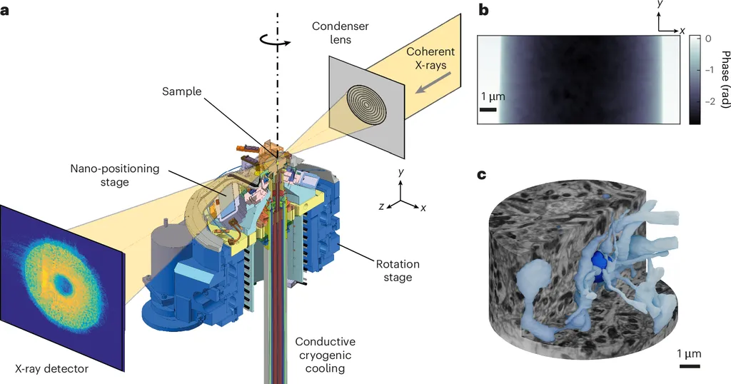
The study, published in the journal Nature Communications, focuses on X-ray ptychography, a coherent diffraction imaging technique that has become a staple at synchrotron facilities for its ability to non-destructively investigate the structure of matter at the nanoscale. Applications range from brain imaging to battery materials, making it a versatile tool for both medical and industrial research.
Until now, the stringent requirements for X-ray beam quality have limited the use of X-ray ptychography to large synchrotron facilities. However, the researchers have shown that the quantitativeness of this technique is preserved even in a laboratory setting, despite the drastic decrease in X-ray flux compared to synchrotron instruments. They achieved this by adapting the method to image a brain tissue phantom, demonstrating the technique’s potential for biological applications.
The team also outlined the current challenges and limitations of using laboratory X-ray sources for quantitative nano-imaging. They highlighted the need for further development and future directions to fully unleash the power of this technique for a broader user community. One of the key challenges is improving the X-ray flux in laboratory settings to enhance the technique’s resolution and speed.
For the energy sector, this development could have practical applications in the imaging and analysis of battery materials and other energy-related nanostructures. By enabling more accessible and non-destructive high-resolution imaging, researchers can better understand the intricate details of these materials, potentially leading to improvements in their performance and efficiency. Additionally, the technique could be applied to investigate the structural changes in materials under different conditions, providing valuable insights for the development of new energy technologies.
In conclusion, the study by Cipiccia and her team represents a significant step towards making advanced imaging techniques more accessible. By demonstrating the feasibility of quantitative phase nano-imaging with laboratory X-ray sources, they open up new possibilities for research in both medical and industrial fields, including the energy sector.
This article is based on research available at arXiv.